PS Capture™ Exosome Flow Cytometry Kit
Features
- High-Sensitive Qualitative Analysis
- Easy Operation by Magnetic Beads
- Direct Detection without Purification
- Total 3 hours ~ from Isolation to Staining ~
Sample Type·Kit Contents
- Cell Culture Supernatant
- Serum
- Plasma (Heparin and EDTA)
| Kit Components (300 assays) | Amount |
|---|---|
| Exosome Capture Beads | 1 x 3 mL |
| Washing Buffer (10×) | 2 x 45 mL |
| Exosome Binding Enhancer (100×) | 1 x 15 mL |
Princeple
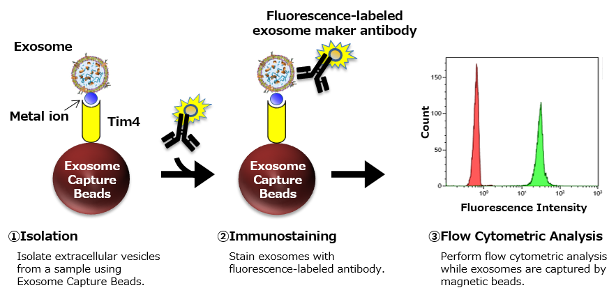
Qualitative analysis of exosomes in cell culture supernatant of K562 cell line
Exosomes in cell culture supernatant of K562 cells were isolated using PS Capture™ Exosome Flow Cytometry Kit or each of anti-CD81-, CD9- and CD63-antibody-immobilized magnetic beads (supplier A), followed by flow cytometric analysis of exosome surface antigens after immunostaining with fluorescence-labeled antibodies.
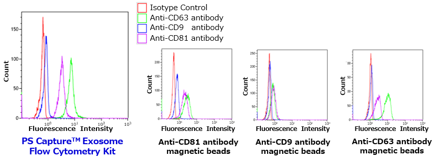
| Sample | Cell culture supernatant of K562 cells: 33 μL/Assay |
|---|---|
| Detection antibody | PE-anti-CD63 (BD Biosciences, 556020) PE-anti-CD9 (Novus Biologicals, NB100-77915PE) PE-anti-CD81 (Novus Biologicals, NBP1-44861PE) |
- PS Capture™ Exosome Flow Cytometry Kit
- Anti-CD81 antibody immobilized magnetic beads
- Anti-CD9 antibody immobilized magnetic beads
- Anti-CD63 antibody immobilized magnetic beads
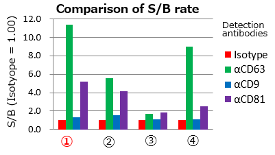
Each signal value was normalized using the value of Isotype.
It was confirmed that the PS Capture™ Exosome FCM Kit can detect the exosome surface antigen with high sensitivity compared with competitors' products, regardless of which detection antibody is used.
Qualitative analysis of exosomes in serum and plasma
Exosomes in human serum and human plasma (EDTA plasma, heparin plasma) were isolated using PS Capture™ Exosome Flow Cytometry Kit, followed by flow cytometric analysis of exosome surface antigens after immunostaining with PE-labeled mouse IgG isotype control and PE-labeled anti-human CD9 antibody.
| Sample: 33 μL/Assay each |
Human serum Human heparin plasma Human EDTA plasma (buffer exchanged) |
|---|---|
| Detection antibody | PE-labeled anti-CD9 antibody (Novus Biologicals, NB100-77915PE) |
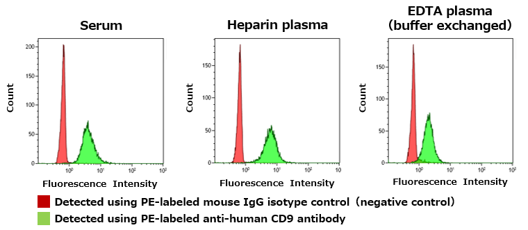
In any of the samples, the peak shift of fluorescence intensity was confirmed when stained with PE-labeled anti-human CD9 antibody.
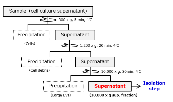
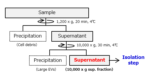
Flowchart of Sample Preparation
Cell culture supernatant
Serum・Heparin plasma
EDTA plasma
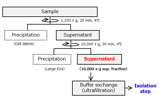
Protocol for buffer exchange (ultrafiltration)
Perform buffer exchange of 1 mL centrifuged EDTA plasma sample with 50 mL of TBS buffer.
- Add 19 mL of TBS to Vivaspin20 (100K).
- Add the 1 mL of centrifuged EDTA plasma sup. to the Vivaspin20 (100K) of 1. and mix (solution A).
- Centrifuge solution A at 4°C. (Refer to centrifugal condition in the manufacturer’s instruction manual.)
- Add 10 mL of TBS to solution A when the upper liquid volume is dropped (solution B).
- Centrifuge solution B at 4°C.
- Add 10 mL of TBS to solution B when the upper liquid volume is dropped (solution C).
- Centrifuge solution C at 4°C.
- Add 11 mL of TBS to solution C when the upper liquid volume is dropped.
- Centrifuge solution C at 4°C until the volume becomes under 1 mL.
- Proceed to “Isolation step”.
In the case of EDTA plasma samples, EDTA inhibits the interaction between EVs and Exosome Capture Beads, thus please perform the buffer exchange beforehand.
Vivaspin20 M.W. cut off: 100K (Sartorius, VS2041) and 50 mL of TBS are required.
Recommended reaction scale
Basic protocol is set as 2 reactions using a 1.5 mL microcentrifuge tube to isolate EVs from samples with Exosome Capture Beads.
For scale-up, increase the amount of Exosome Capture Beads and samples. Recommended reaction scale is presented in the right table.
Note: 10 reactions are maximum for one 1.5 mL microcentrifuge tube.
| Qty of Reaction | Exosome Capture Beads (µL) | Sample Volume(µL) |
|---|---|---|
| 2 reactions (basic) |
30 | 100 |
| 3 reactions | 40 | 133 |
| 4 reactions | 50 | 167 |
| 5 reactions | 60 | 200 |
| 6 reactions | 70 | 233 |
| 7 reactions | 80 | 267 |
| 8 reactions | 90 | 300 |
| 9 reactions | 100 | 333 |
| 10 reactions | 110 | 367 |
Outline of Procedure ~Basic Protocol for 2 reactions~
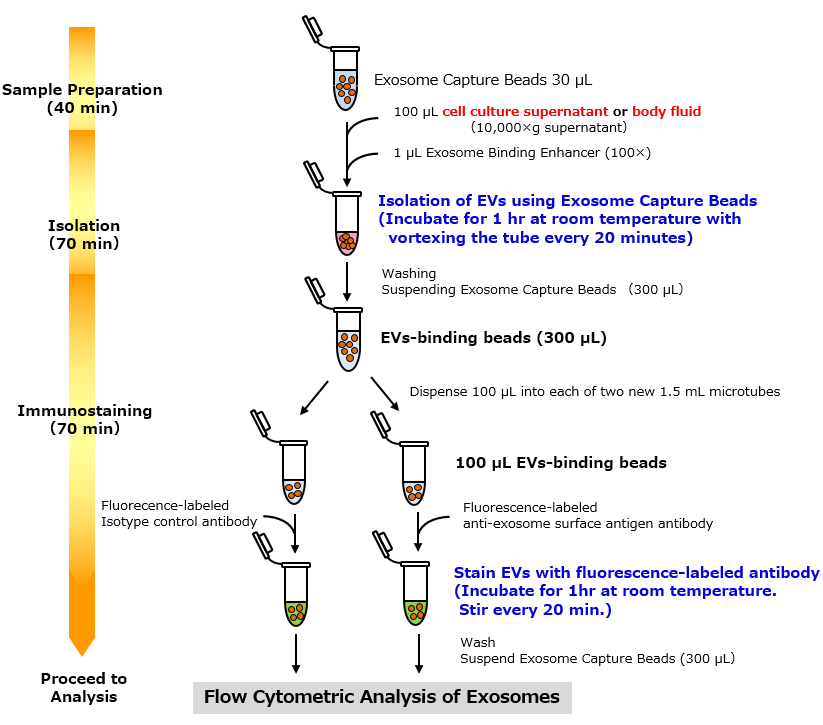
The kit can detect any surface marker proteins on extracellular vesicles (EVs) with high sensitivity by flow cytometry after capturing EVs by PS Affinity Method※1. Before use, it is necessary to prepare fluorescence-labeled primary antibody against exosome surface marker protein, or primary antibody and fluorescence-labeled secondary antibody.
Reference
- "A novel affinity-based method for the isolation of highly purified extracellular vesicles", W. Nakai, T. Yoshida, D. Diez, Y. Miyatake, T. Nishibu, N. Imawaka, K. Naruse, Y. Sadamura & R. Hanayama, Sci Rep 6, 33935 (2016).