![]()
This product, called Cellcampus®, is a preparation of type I collagen derived from the scales of farm-raised tilapia, a tropical fish. Cellcampus® is manufactured by Taki Chemical Co., Ltd. in Japan. 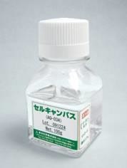
There are no known viruses transmitted from tilapia to humans. Furthermore, tropical fish collagens show higher denaturation temperatures, as compared with collagens derived from non-tropical fish. Thus, Cellcampus® is perfect for tissue and cell culture applications in the field of regenerative medicine. 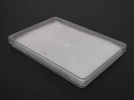
It is well documented that tilapia scale collagen exhibits an excellent fibril-forming ability. Moreover, this collagen can enhance cell proliferation. Therefore, Cellcampus® is also ideal for general cell culture protocols.
Features
- Safe and Reliable Fish-Derived Product
There is no known fish viruses infect to humans. Thus, fish collagen is a not human health risk. - Highly thermostability
The source of type I collagen is a farm-raised tropical fish (tilapia). The tropical fish (tilapia) collagen has a high denaturation temperature, making it functionally active at typical cell culture temperatures. (Cellcampus® is available for coating cell culture dish surface.) - Outstanding Fibril-Forming Ability (see Ref. 1)
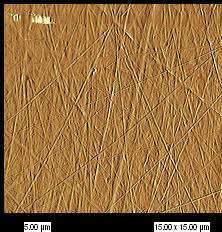
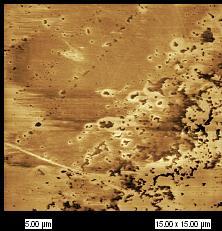
Left:Cellcampus® (Collagen isolated from fish scales) Right:Collagen isolated from pig skin
culture dishes were coated with the indicated reagents and subsequently air-dried at room temperature. These atomic force microscopy images show the surface characteristics of the culture dishes.
Fish collagen extracted from tilapia scales has an increased ability to reassemble into its physiological form (i.e., a fibrillar structure with systematically arranged collagen molecules). Thus, Cellcampus® allows you to coat culture dish surfaces with native collagen fibrils.
Overview
-
Cellcampus® AO-03A(Liquid)
Quality Control Testing Acceptance Criteria Appearance Clear and colorless solution Collagen concentration (%) 0.30~0.36 Specific optical rotation (°) -350~-450 pH 3.0~5.0 Bioburden Undetectable Heavy metals(ppm) ≤ 20 (not exceeding the detection limit) Arsenic(ppm) ≤ 2 (not exceeding the detection limit) -
Cellcampus® FD-08G(Lyophilized)
Quality Control Testing Acceptance Criteria Appearance White sponges Weight(g) 0.80~0.90
Application 1
Improved Cell Proliferation (Ref. 2)
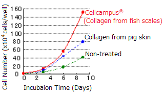
Growth Curve of HeLa Cells
Cellcampus® is effective in promoting cell growth※ 。
- A similar effect was observed with the cell lines L929, MC3T3, and Saos-2.
- Cellcampus® can also facilitate the proliferation of cells cultured in low-serum media
Application 2
Enhanced Cell Differentiation (Ref. 3)
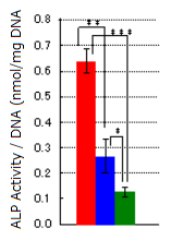
*P<0.05, **P<0.005, ***P<0.0001 |
[Cell Culture and Assay Methods] |
Alkaline phosphatase (ALP) activities were determined after inducing osteoblast differentiation in human mesenchymal stem cells. This result indicates that Cellcampus® can promote the differentiation of these stem cells※.
- We also confirmed that Cellcampus® can elevate the expression of the early osteoblastic marker genes BMP2 and OPN
References
- J. Tanaka,et al. BioIndustry, 26(8), 26-32(2009).
- Y. Imaizumi, et al. MaterialsIntegration, Vol.23(2), 27-31(2010).
- R. Matsumoto, et al. BioIndustry, 28(11), 22-26(2011).